Research

Raman Spectroscopy

Reading the molecular fingerprint of living systems — from single-cell gene expression to needle-free sensing through the skin.

Overview

Raman spectroscopy reads a sample's molecular vibrations directly, label-free and non-destructively. The lab pushes it in two directions: using machine learning to turn Raman spectra of living cells into single-cell gene-expression readouts, and miniaturizing Raman into compact sensors that read chemistry straight through the skin. Together they open biomedical applications across diabetes, aging, cancer, and image-guided surgery.

Technology Directions

New directions we're pushing

Two active fronts — one computational, one instrumental — each extending what a Raman spectrum can tell us about living systems.

DIRECTION 01

Raman2RNA — reading gene expression from light

Single-cell RNA sequencing reveals a cell's genetic program, but it destroys the cell — a snapshot, never a movie. Raman2RNA (R2R) infers genome-wide single-cell expression from label-free Raman microscopy instead. A full Raman spectrum is acquired at every pixel, and a neural network trained against single-molecule FISH and single-cell RNA-seq maps each living cell's spectral fingerprint into gene-expression space. Because Raman is non-perturbing, the same living cells can be read repeatedly over time: R2R followed expression dynamics through mouse embryonic-stem-cell differentiation and iPSC reprogramming, resolving where lineages diverge earlier than destructive snapshot methods could.
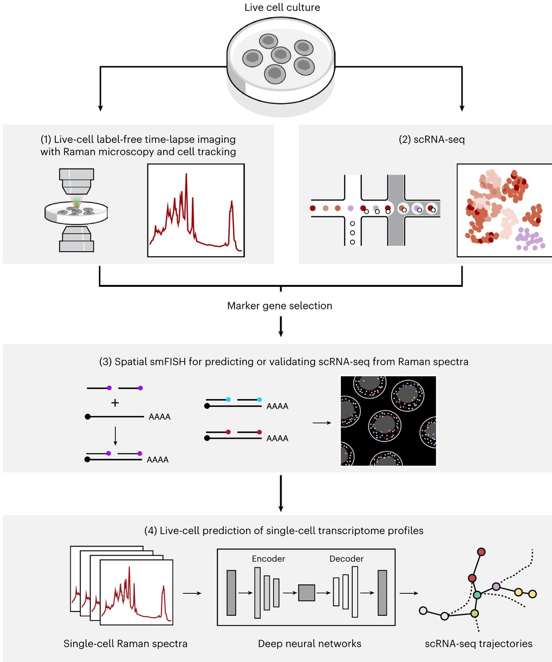
Raman2RNA method
Instrument:Living cells are imaged by hyperspectral Raman microscopy; a deep neural network trained against smFISH and single-cell RNA-seq maps each cell's spectrum to a genome-wide expression profile and trajectory.Kobayashi-Kirschvink et al. · Nature Biotechnology 2024
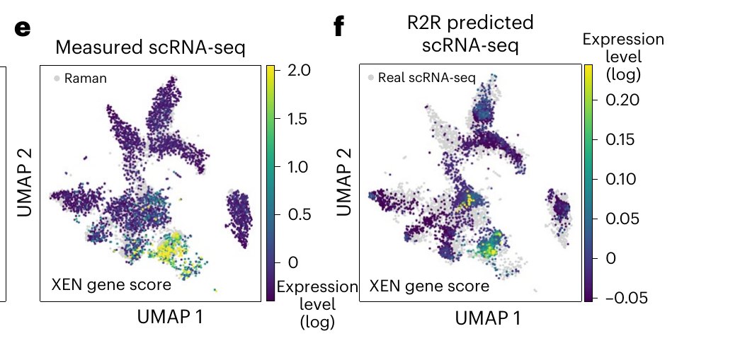
Raman2RNA prediction
Demonstration:R2R-predicted single-cell expression of a stem-cell lineage marker (right) reproduces the measured single-cell RNA-seq pattern (left) — inferred from the Raman signal alone.Kobayashi-Kirschvink et al. · Nature Biotechnology 2024
DIRECTION 02

Non-invasive optical glucose sensing

For people with diabetes, the standard glucose measurement is still a finger-stick blood draw — accurate, but painful and invasive enough that many test less often than they should. Minimally invasive continuous monitors (Dexcom, Abbott) have transformed daily management, but they still insert a microneedle filament under the skin, must be replaced every couple of weeks, and remain costly. A truly non-invasive optical reading could improve on both — if Raman can genuinely detect glucose through skin. The lab answered that question directly: with an off-axis fiber-optic Raman system it recorded the first direct observation of glucose's own Raman fingerprint in living tissue, tracking blood-glucose swings in vivo. Building on that proof, a band-pass Raman design reads just three narrow Raman bands — the glucose peak near 1125 cm⁻¹ and two references — shrinking the optics toward a compact, potentially wearable sensor.
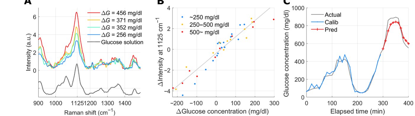
Direct glucose Raman fingerprint in vivo
Proof of principle:The first direct observation of glucose's own Raman fingerprint in living tissue — the 1125 cm⁻¹ glucose peak grows with concentration (left), scales linearly with blood glucose (center), and tracks it in vivo (right; R = 0.95).Kang et al. · Science Advances 2020
Band-pass Raman device
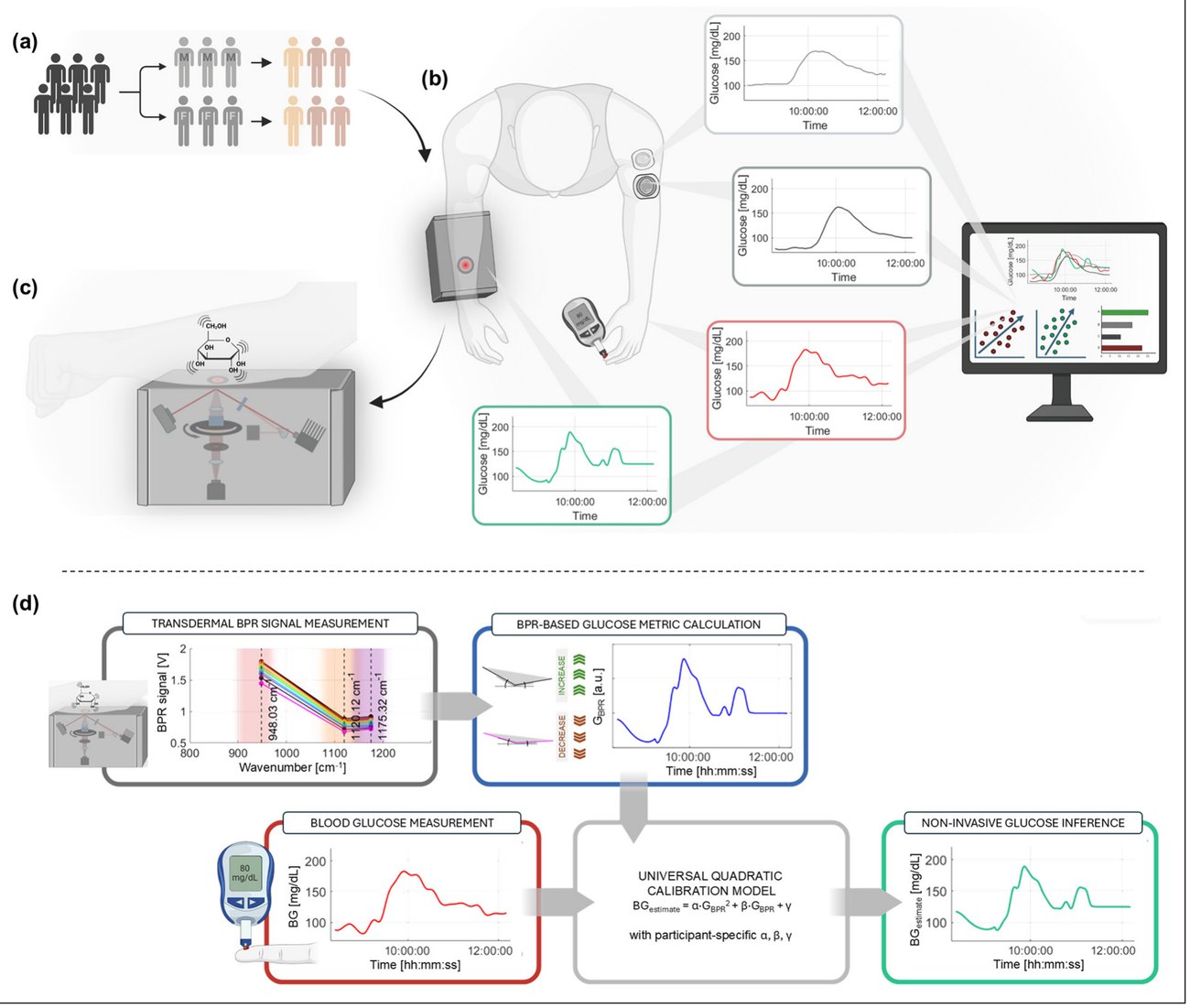
Next-generation device:A compact band-pass Raman monitor reads three narrow Raman bands through the skin with an 830 nm laser — no needle. In a six-participant human trial it matched commercial CGMs (mean absolute relative difference ~11%), a step toward a wearable optical sensor.Bresci et al. · J. Diabetes Science & Technology 2026
Biomedical Applications

Putting the technology to work

Biomedical questions the lab takes on with its Raman imaging and sensing assets — spanning metabolic disease, aging, cancer, and the operating room.
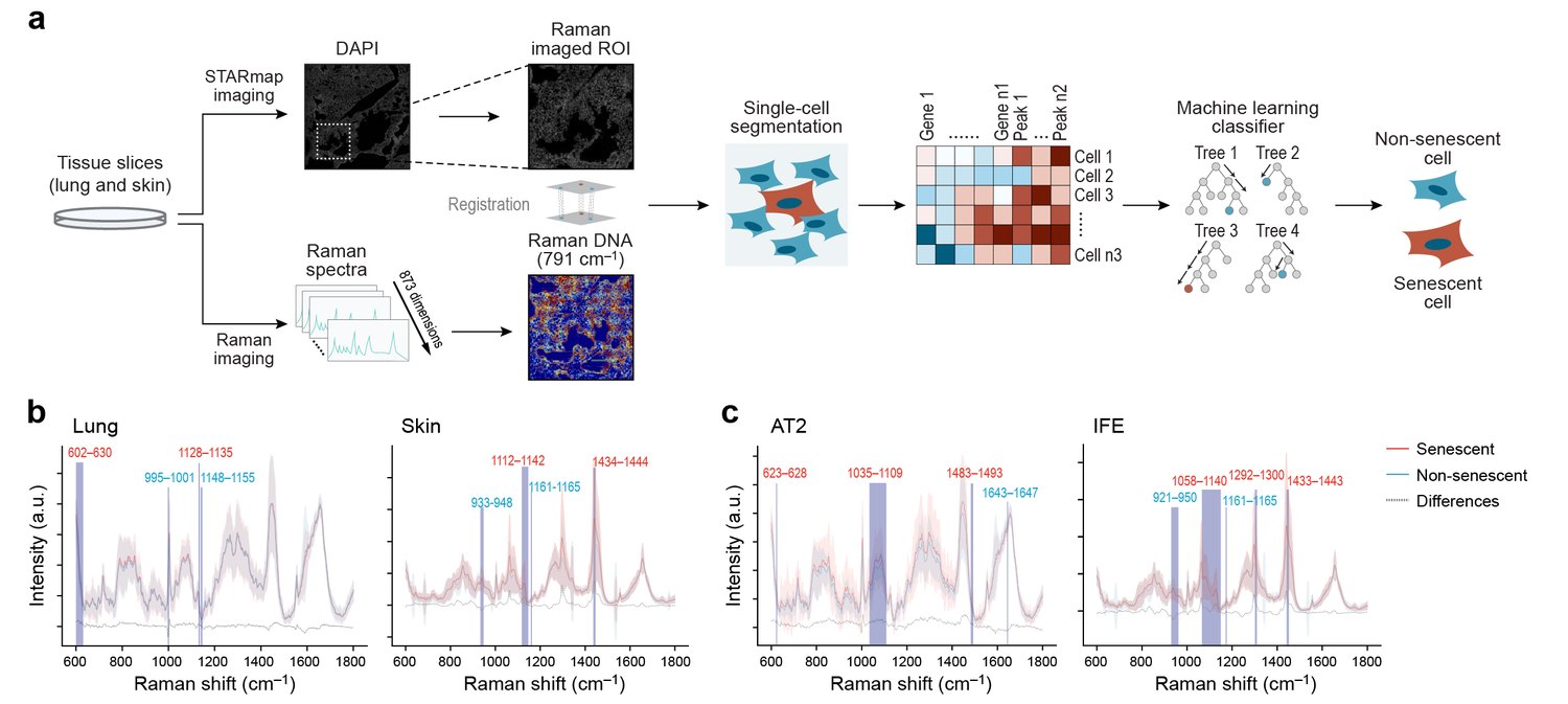
Cellular senescence detection

Cellular senescence & aging

Senescent cells drive aging but lack a single marker. Pairing label-free Raman signatures with machine learning and spatial transcriptomics (RamanOmics), the lab links a reproducible lipid-associated Raman band to senescent (p21⁺) cells in aged lung and skin — biochemical information that sharpens senescence detection beyond gene expression alone.Zhang et al. · Nature Aging (in press) 2026
Label-free cancer-cell phenotyping

Label-free cancer-cell phenotyping

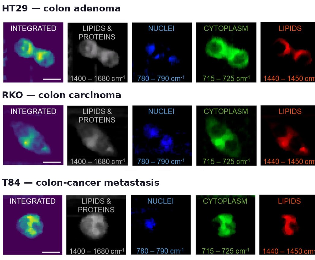
Combining Raman molecular fingerprints with 3D quantitative phase tomography, living colon-cancer cells are characterized without stains or fixation — distinguishing adenoma, carcinoma, and metastatic lines by their combined morpho-molecular signatures.Bresci et al. · Communications Biology 2024
Monitoring cancer treatment response

Monitoring cancer treatment response

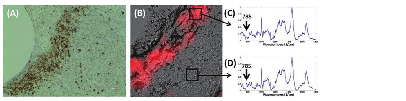
Knowing whether a chemotherapy is working can take weeks of waiting for a tumor to shrink. Raman detects the molecular signature of drug-induced cell death directly in tumor tissue — a sharp drop in the DNA band as doxorubicin takes effect — within 24 hours, mapping responding regions in spatial agreement with apoptosis (cleaved-caspase) histology.Jonas, Kang et al. · Analyst 2018
Raman-guided needle placement

Image-guided needle placement

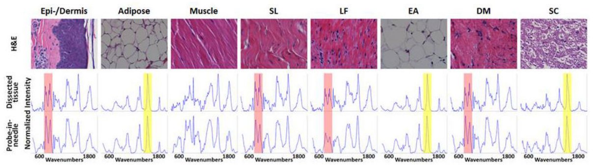
A Raman probe inside an epidural needle identifies each tissue layer from skin to spinal cord by its molecular signature — collagen-rich ligaments (939 cm⁻¹) versus lipid-rich fat and cord (1450 cm⁻¹) — giving real-time feedback on needle-tip location for safer neuraxial placement.Anderson, Kang et al. · Anesthesiology 2016