
Research

Interferometric Microscopy

Reading the phase of light to measure the shape, density, and motion of living cells — label-free, in three dimensions.

Overview

Interferometric microscopy reads the phase of light — the minute delay a specimen imposes on a light wave — to image cells and tissues label-free, quantifying their three-dimensional shape, refractive index, and nanometer-scale motion with no stains. The lab pushes two directions: bringing temporal focusing into phase imaging for scanless, high-speed 3D quantitative phase microscopy, and a structured-illumination scheme (dSIM) for fast, large-field volumetric refractive-index imaging; a single-frame tomographic phase cytometer (xSCYTE) extends the toolkit to high throughput. These methods reach applications from single-cell blood diagnostics to the biophysics of biological nanostructures and nuclear mechanics.

Technology Directions

New directions we're pushing

Two ways to make quantitative phase imaging faster and reach further into three dimensions — one using temporal focusing, one using structured illumination.

DIRECTION 01

TF-QPM — scanless 3D quantitative phase microscopy

Quantitative phase microscopy turns the tiny delays a cell imposes on light into a label-free map of its structure — but building a depth-resolved 3D image usually means scanning, which is slow. TF-QPM brings temporal focusing — a scanless, whole-field way to select a single depth plane, until now used only for fluorescence — into coherent phase imaging for the first time. A spectrally chirped pulse recompresses to its shortest duration only at the focal plane, so only that plane interferes with the reference while everything above and below decorrelates. The result is single-shot optical sectioning whose thickness is set by the objective (~0.9 µm) rather than the light source, at camera-limited rates up to ~3.7 kHz — about an order of magnitude faster than prior full-field 3D phase microscopy — with ~2 nm axial sensitivity.
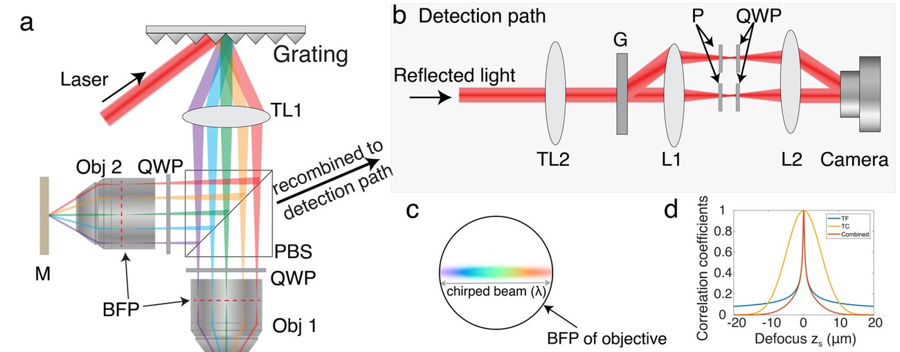
TF-QPM setup
Instrument:A Linnik interferometer with temporally-focused illumination: the chirped pulse recompresses only at the focal plane, giving scanless optical sectioning, while off-axis holography recovers the full complex field in a single shot.Lin et al. · LBRC (in preparation)
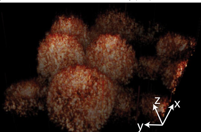
3D label-free cell imaging
Demonstration:Volumetric, label-free rendering of cells reconstructed from single-shot 3D quantitative phase imaging — resolving nuclei, membranes, and fine features with no stain.Lin et al. · LBRC (in preparation)
DIRECTION 02

dSIM — large-field, high-speed 3D refractive-index imaging

Two established 3D phase methods force a trade-off: tomographic phase microscopy is fast but sees only a small field, while dark-field Fourier ptychography covers a large field but is slow (~1 volume/s), with weak, noisy signals and hours-long reconstructions. dSIM (differential structured illumination microscopy) is designed to get both. Two coherent beams — one at a bright-field angle, one at a steep dark-field angle beyond the objective's collection cone — interfere on the sample; the objective rejects the direct dark-field beam, but its weak high-angle scattering rides on the strong bright-field background through heterodyne gain. Subtracting a bright-field-only image isolates that dark-field signal and keeps the object-to-intensity relationship linear, enabling fast, closed-form 3D refractive-index reconstruction over a millimeter-scale field — targeting ~100 volumes/s and 3D image cytometry beyond half a million cells per second.
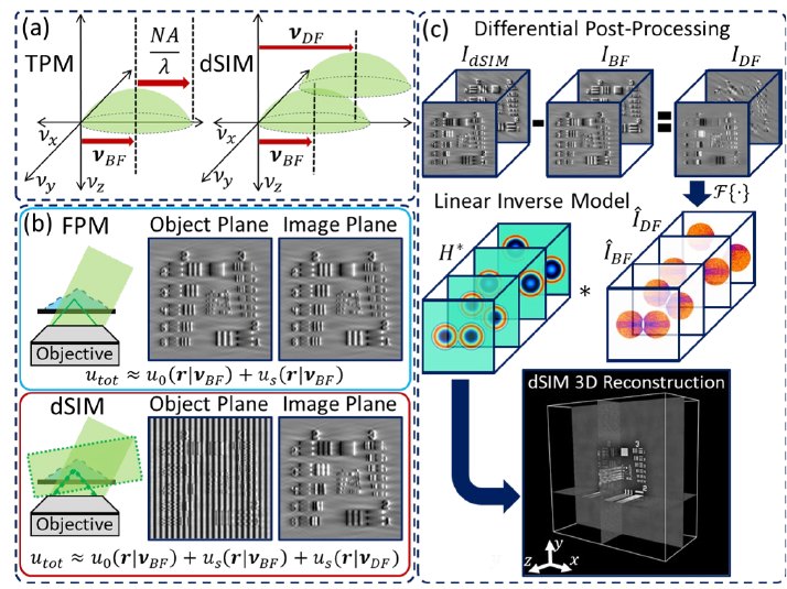
dSIM concept
Concept:dSIM encodes high-angle dark-field scattering into bright-field-like images through heterodyne gain; a differential subtraction and a linear inverse model then reconstruct 3D refractive index.So et al. · LBRC (proposed)
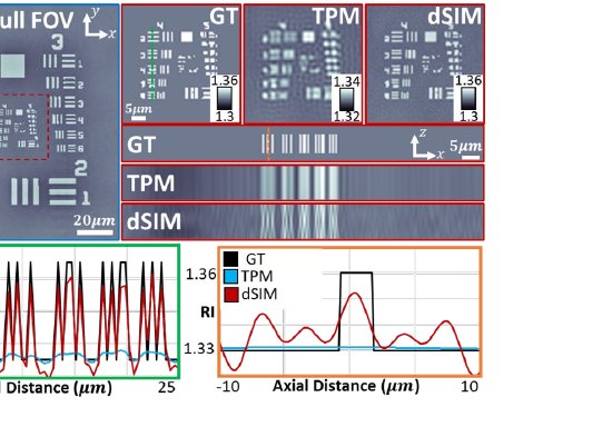
dSIM simulation
Simulation:In simulation, dSIM recovers a sub-resolution phase target with sharper lateral and axial resolution and higher refractive-index contrast than tomographic phase microscopy (TPM). Instrument in development.So et al. · LBRC (proposed)
Biomedical Applications

Putting the technology to work

Where the lab's phase and interferometric imaging assets meet biology — from single-cell blood diagnostics to the physics of biological nanostructures and the mechanics of the cell nucleus.
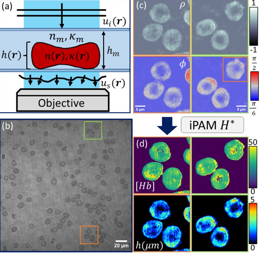
iPAM single-cell overview
Vaso-occlusive-crisis risk prediction

Single-cell sickle-cell analysis

Standard blood counts report only bulk averages, missing the cell-to-cell variation that may drive sickle-cell crises. iPAM measures each red blood cell's quantitative phase (shape, volume) and optical absorption (hemoglobin) at once, label-free and at high throughput — recovering bulk clinical metrics to within ~5% (left). Beyond diagnosis, single-cell sub-populations invisible to standard counts carry predictive signal: a median-height variation measure separates patients by vaso-occlusive-crisis risk (right; AUC 0.77).Matlock et al. · LBRC (submitted)
Nuclear envelope mechanics

Nuclear-envelope mechanics

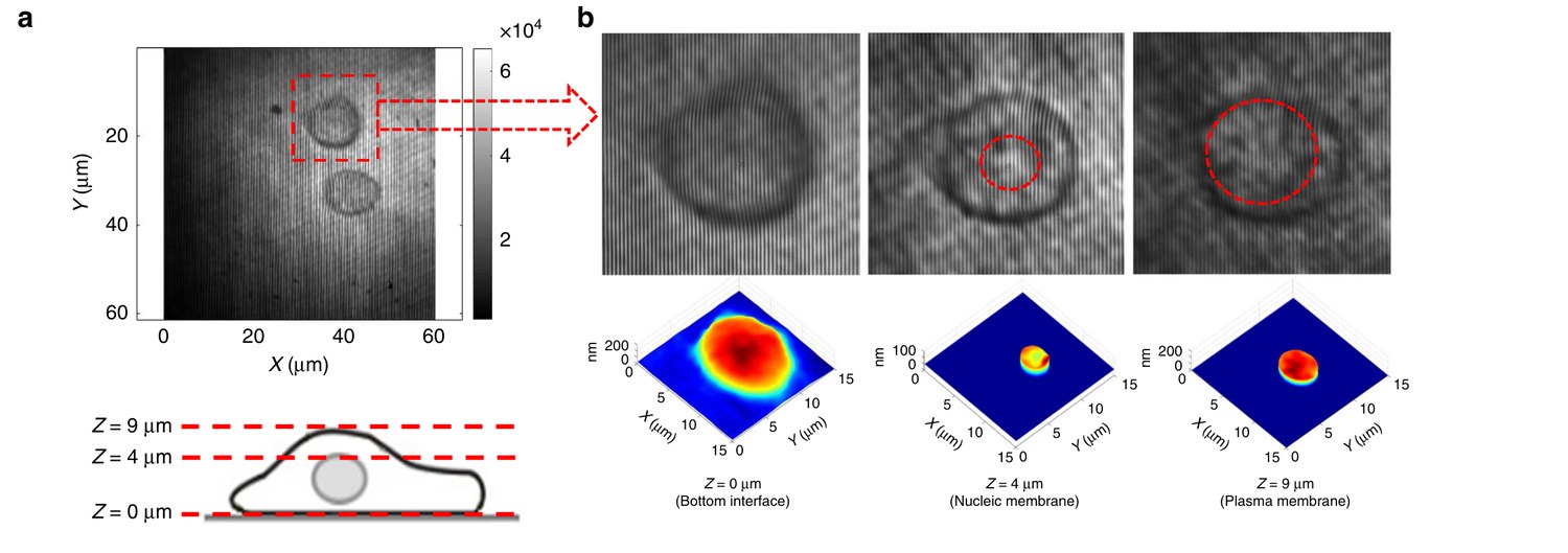
Nuclear mechanics report on cell state and disease, from stem-cell differentiation to cancer metastasis. A depth-resolved confocal reflectance interferometric microscope measures nanometer-scale membrane fluctuations separately at the plasma membrane and the nuclear envelope of living cells — label-free and non-contact — showing the nuclear envelope's greater stiffness and its response to substrate rigidity.Singh et al. · Nature Communications 2019
Butterfly scale development timeline
Ridge-formation mechanics

Butterfly wing-scale development

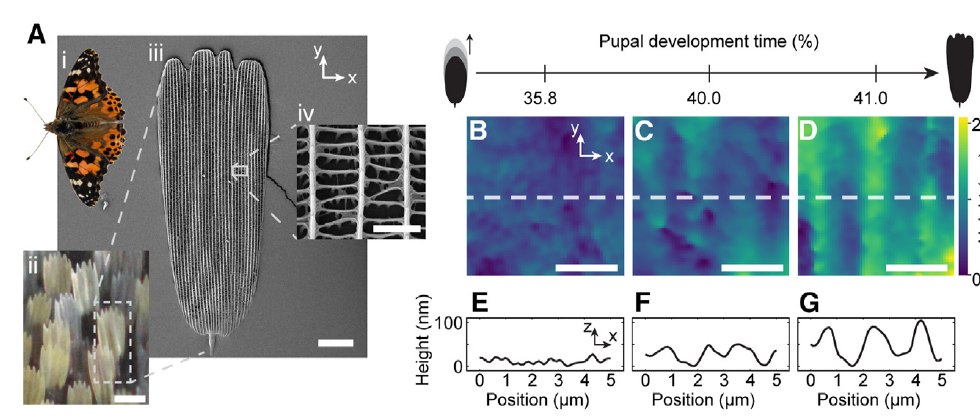
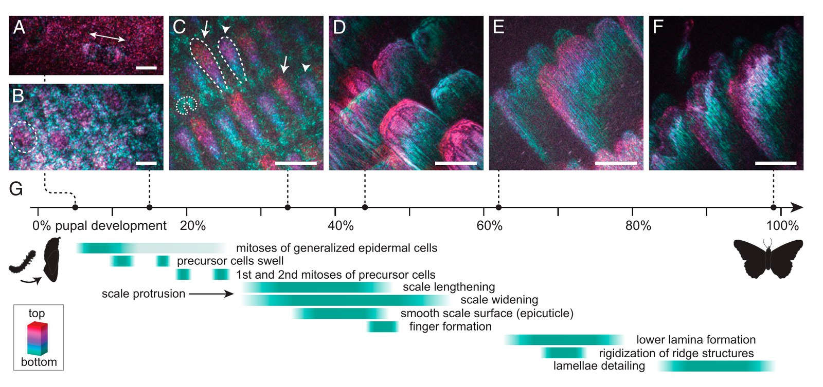
Butterfly wing scales self-assemble intricate light-manipulating nanostructures — a model for biological photonic fabrication. Through a window in the living chrysalis, label-free interferometric phase imaging captured the continuous in-vivo development of individual scale cells across the ~10-day pupal stage, from epithelial sheet to ridged, light-scattering scale (left). The same phase data revealed the mechanism of the earliest ridges: the growing scale membrane, confined by regularly spaced actin bundles, buckles into regular protoridges (right).McDougal et al. · PNAS 2021 · Totz et al. · Cell Rep. Phys. Sci. 2024