
Seeing living tissue in molecular detail — deeper and faster through the fog of scattering.
Multiphoton fluorescence has anchored the LBRC's imaging science for decades. Two threads run through the work today. We push new technology directions that extend what fluorescence-based imaging can see — computationally recovering wide-field images from light that tissue scattering would otherwise scramble, and pulling endogenous metabolic signals out of deep tissue by acoustic detection, where scattering and absorption would otherwise keep them from any optical measurement. And we turn the lab's imaging assets into biomedical applications, from human neurodevelopment and cortical function to cancer-linked mutations and tissue fibrosis.
Two active fronts in fluorescence and multiphoton imaging — one computational, one physical — each aimed at seeing further into living tissue than fluorescence detection alone allows.
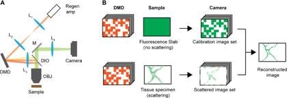
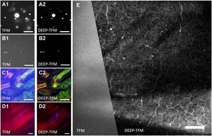
Temporal-focusing microscopy excites and images an entire field of view at once, rather than scanning one point at a time — which makes it fast. Its weakness is depth: the fluorescence it generates is scattered on the way back to the camera, so the wide-field image blurs just a few scattering lengths into tissue. DEEP (De-scattering with Excitation Patterning) keeps the speed of wide-field temporal focusing while undoing that scattering computationally. A digital micromirror device (DMD) imprints a sequence of patterned excitations onto the temporally-focused beam; because the patterns are carried by long near-infrared wavelengths they hold their shape deep in tissue, and the camera records the scrambled wide-field emission for each one. The same patterns are first recorded on a thin, non-scattering fluorescent slab to calibrate the system, and the de-scattered image is then reconstructed from the tissue measurements.
The reconstruction is what makes it efficient. Because wide-field detection already preserves the low-spatial-frequency content and the algorithm exploits the image's compressibility — a compressive-sensing / wavelet-sparsity prior — only a few hundred patterns are needed, versus the millions of measurements a point scan of the same volume would take, and the acquisition time does not grow with field of view. DEEP² pushes the computation further: a deep neural network (a U-Net with attention) learns the inverse mapping directly. Because matched training pairs are nearly impossible to collect in living tissue, the network is trained entirely on realistic DEEP images synthesized by a physics-based forward model from existing point-scanning data — cutting the patterns needed roughly eight-fold, to about 32. Both have been demonstrated from bead phantoms and excised tissue through to in-vivo mouse cortex.
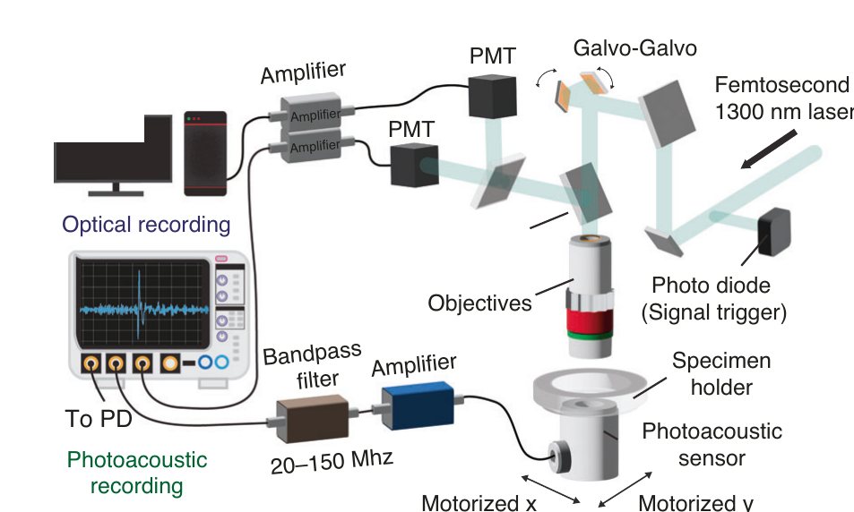
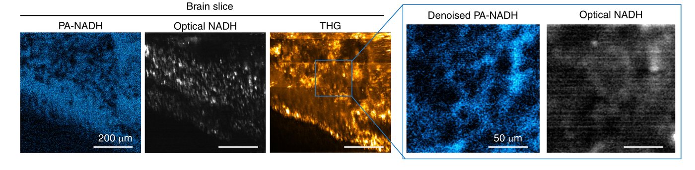
Fluorescence readout of the metabolic coenzyme NAD(P)H — nicotinamide adenine dinucleotide (NADH) and its phosphorylated form, nicotinamide adenine dinucleotide phosphate (NADPH), which optical imaging cannot separate — is capped at a few hundred microns, because its blue emission is strongly absorbed and scattered by tissue. This direction reads the same molecule a different way. A 1300 nm femtosecond laser drives three-photon absorption in endogenous NAD(P)H; because NAD(P)H is a poor emitter, most of that absorbed energy turns to heat — a tiny thermoelastic expansion that launches an ultrasound pulse, detected acoustically. Ultrasound leaves tissue far more easily than blue light, so this label-free metabolic signal can be followed much deeper: NAD(P)H was detected to roughly 700 µm in brain slices and about 1.1 mm in cerebral organoids — several times the optical limit — at single-cell resolution.
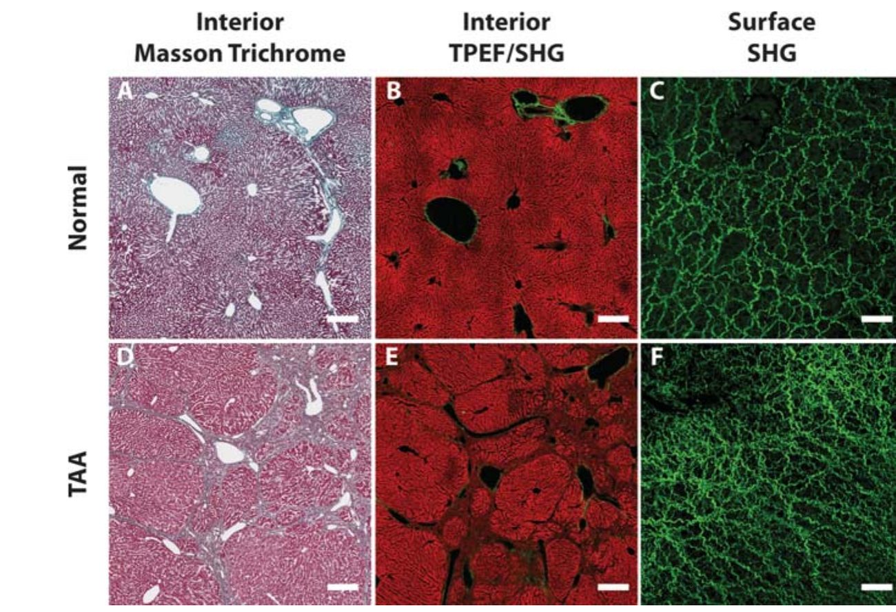
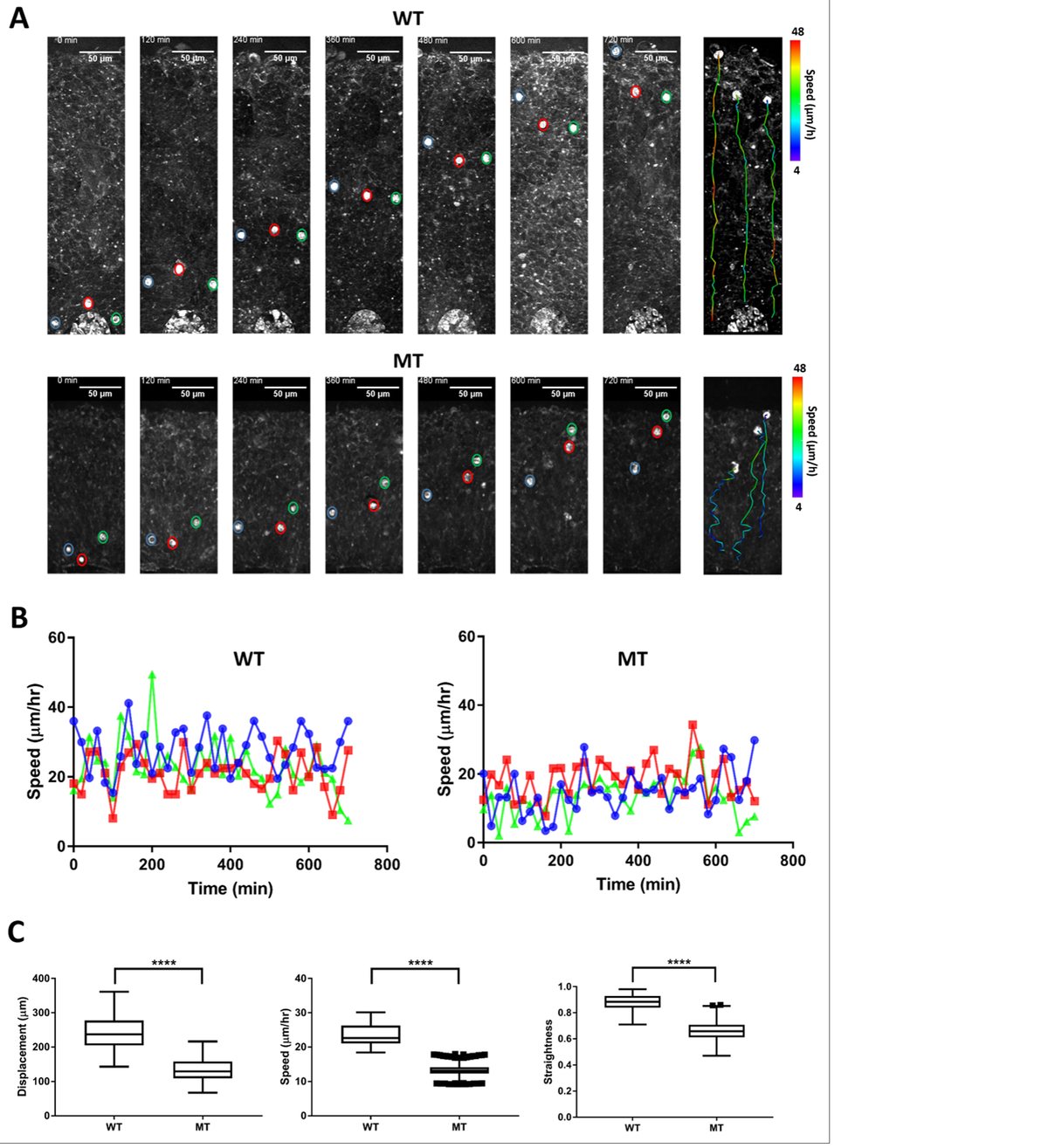
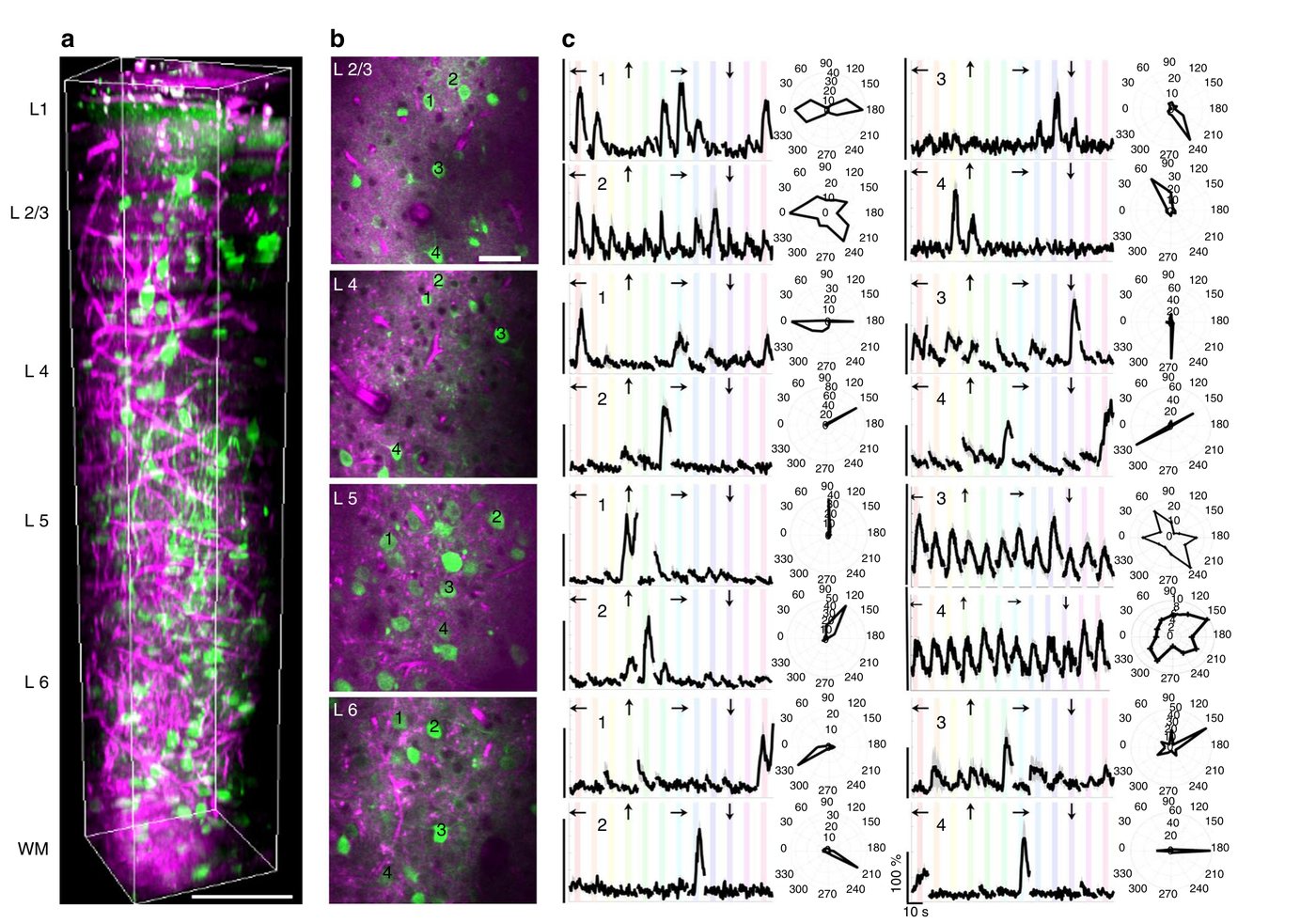
Biomedical questions the lab takes on by applying its fluorescence and multiphoton imaging assets — with our collaborators — across cancer biology, clinical pathology, and neuroscience.